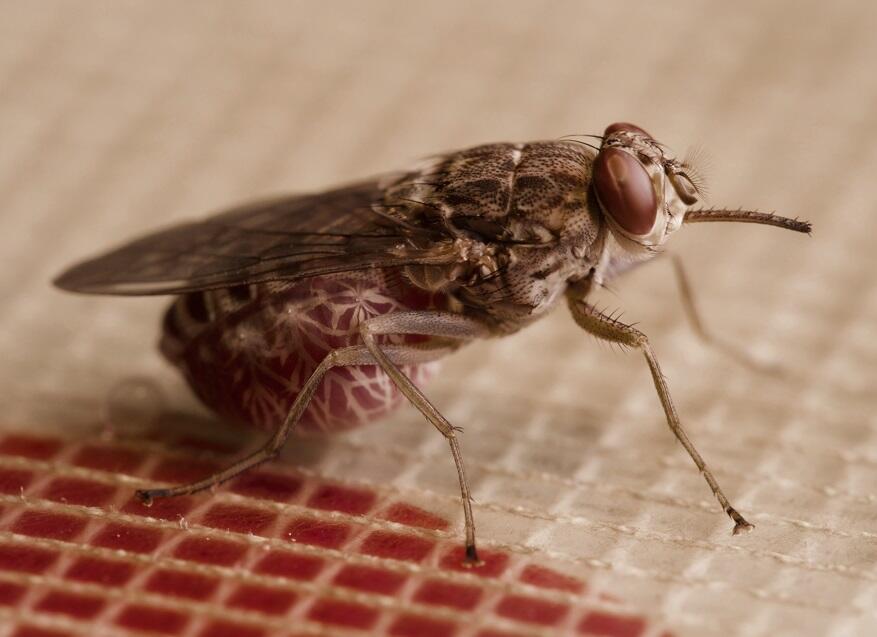
An LSTM led study on how the deadly trypanosome parasite colonizes tsetse flies has been published by Nature Microbiology. The research was led by Dr Alvaro Acosta-Serrano from LSTM’s Department of Vector Biology with colleagues Dr Clair Rose, Dr Aitor Casas-Sánchez and Dr Naomi Dyer as shared first authors. The study also involved colleagues from the German University of Würzburg and the University of Liverpool.
These blood-borne trypanosome parasites are transmitted by tsetse flies and cause potentially fatal disease in humans (trypanosomiasis, also known as sleeping sickness) and livestock (Nagana) in sub-Saharan Africa.
The key step to trypanosome transmission, following ingestion of the parasite-infected mammalian blood, is the establishment of an infection within the gut of the tsetse fly. The exact location where the parasite “hides” –known as the ectoperitrophic space (ES) – is located between the gut epithelium and the peritrophic matrix (PM). The PM is the insect equivalent to the mucous layer that protects the mammalian digestive system, but rather than mucous, it is a filmy sleeve that surrounds the incoming bloodmeal. The parasites must multiply within the ES to safely continue through all the development stages before they can migrate to the fly’s salivary glands. Only when they colonise the salivary glands can the parasites be released into the fly saliva, ready to be transmitted when the fly takes its next blood meal. The authors decided to investigate an overlooked, but fundamental, biological question: how do the parasites reach the ES in the first place, evade the fly’s immune system and impact disease transmission?
To address this question, the team used a combination of state-of-the-art confocal laser scanning microscopy and electron microscopy to visualise trypanosome parasites within the fly gut. These enabled them to obtain striking 3D-images of the precise location of the parasites during the first days of arriving with the blood meal to better understand the dynamics of an early trypanosome invasion within specific tsetse tissues.
The team proved that trypanosomes enter the ES by crossing the newly secreted PM in the proventriculus (PV) – a mushroom-shaped organ that continually synthesises the protective PM to surround the incoming blood meal. This route contrasts with previous beliefs adopted more than forty years ago, and this work now fills an important knowledge gap in trypanosome migration processes in tsetse.
Alongside this discovery, the authors also observed that early PV colonisation leads to a greater chance of establishing salivary gland infections. This process is enhanced by factors present in serum from trypanosome-infected mammalian blood. The exact nature of these serum factors is yet to be elucidated; however, understanding these basic mechanisms could lead to a new tool for disease control.
Dr Álvaro Acosta-Serrano said “I am very pleased with the discoveries reported in our paper, which represents a paradigm shift regarding the way trypanosomes colonize the tsetse fly vector. These findings expand our knowledge on the fascinating interactions between pathogens and disease vectors, and could lead the discovery of new targets to block the transmission of vector-borne pathogens. This work would not have been possible without access to the unique LSTM’s tsetse colony and category 3 confocal facility, the hard work and dedication of a team of talented PhD students and postdocs, and the participation of great collaborators.”
Trypanosoma brucei colonizes the tsetse gut via an immature peritrophic matrix in the proventriculus as published in Nature Microbiology, 20 April 2020.