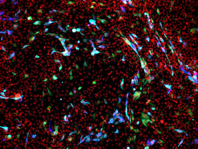
LSTM researcher, Dr Aitor Casas-Sanchez, has filmed SARS-CoV-2 (see below for a clip), the virus which causes COVID-19, whilst it infects human lung epithelial cells and other cell types, as an infection model for COVID-19 research. Importantly this tool will lead researchers to new important discoveries, so we better understand COVID-19 infection dynamics.
LSTM is one of the few sites where this live imaging microscopy can take place, as it has specialised confocal laser scanning microscopes situated within the Category 3 Laboratory whereby infection experiments can be performed to the appropriate biosafety standards whilst imaging the cells for hours or even days.
The team spent more than a year developing the methodology to image the virus, as they had to first modify the virus genetically for it to produce fluorescence which could be visualised under the confocal laser scanning microscope. A special licence had to be obtained by the team to genetically modify SARS-CoV-2.
Dr Casas-Sanchez said: “It took a long time to organise and optimise the methods for visualising the COVID-19 virus under the microscope, especially the process of obtaining a licence for modifying of SARS-CoV-2. However, it was worth the wait as this unique setup is an incredibly powerful tool for studying SARS-CoV-2 and will take our research to a whole new level.”
Prior to the optimisation of this visualisation methodology, Dr Sanchez would have to end an experiment at any specific time point to analyse the results limiting many outcomes including understanding the infection process in its entirety. However, now the team can run experiments in real-time and analyse as the infections are currently taking place.
Dr Casas-Sanchez continued: “Being able to understand how things happen in real-time in such a dynamic way, instead of looking at a few snapshots in time, is incredibly useful.”
Another important aspect of the tool Dr Casas-Sanchez developed included the ability to genetically modify SARS-CoV-2. As the genome of SARS-CoV-2 is RNA-based, and we can only genetically modify DNA, the team had to work with a large DNA molecule that encoded the whole virus genome. These large DNA molecules called infectious clones can be modified and then converted to real viruses. The power of using such infectious clones is that the team can now alter the genome of SARS-CoV-2 with relative ease such as inserting mutations and adding fluorescence markers for example. Such mutant viruses can be used in experiments to understand the dynamics of infection and how new variants arise within populations. At present to study new variants, we need to wait for the incidence of variants occurring in the population to increase before clinical scientists isolate and distribute them which is a time-consuming and logistically- demanding process that can take many weeks. Using infectious clones will mitigate this and speed up our understanding of COVID-19 infections.
The combination of producing infectious clones of SARS-CoV-2 and the live imaging of the infection process is a game-changing tool for better understanding COVID-19 infections. Dr Casas-Sanchez will now look at the effects of sugar molecules called glycans on the surface of SARS-CoV-2 using these modification and live imaging tools. He aims to modify virus to deplete individual glycans to understand their role in the infection process and design molecular tools to target such essential glycans to fight the virus.
Below: Live microscopy over 18 hours of cultured cells inoculated with genetically-modified SARS-CoV-2 infectious clones. Infected cells can be identified over time due to the expression of a red fluorescent protein.